What role does ultrasound play during pregnancy, and is it dangerous for the fetus?
During pregnancy, a woman’s belly shape may confirm pregnancy, but in the 21st century, fetal ultrasound gives us all the other essential information -such as the fetus’s location, weight, development, the overall condition of the pregnant woman, gender, expected delivery date, and more.
Based on ultrasound examination, we can anticipate complications in pregnancy, reduce risks, and increase the chances of a live birth.
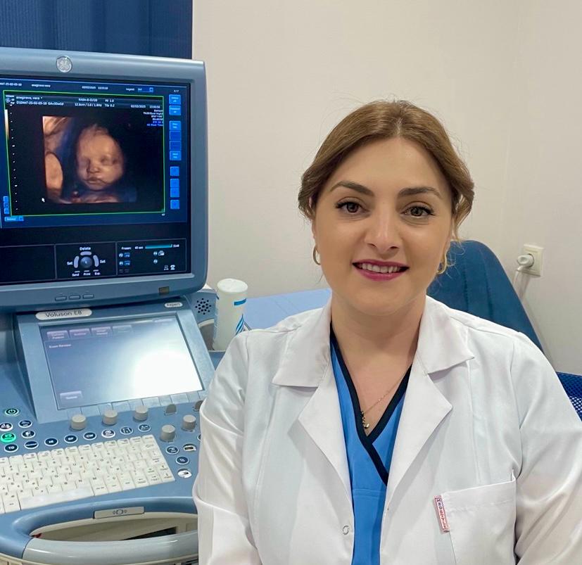
Dr. Tamta Motsonelidze, a radiologist at the Georgian-German Reproductive Center (GGRC), explains the kind of important information we can gather during specific stages of pregnancy and why scheduled and consistent ultrasound monitoring is necessary.
Ultrasound is an examination method that can be performed throughout the entire pregnancy without causing any harm to either the mother or the fetus. It allows us to monitor fetal growth and development, keep an eye on progress, and identify potential issues if any arise.
At the beginning of pregnancy, we need to assess whether the pregnancy is located in the uterus or is ectopic (outside the uterus). If the gestational sac is within the uterus, we must identify the number of sacs. In the case of a twin pregnancy, both the patient and the gynecologist must be informed early on. We also examine the uterus, ovaries, and the entire pelvic area to check for any pathological conditions that might interfere with the pregnancy. At this stage, we often use a transvaginal approach, which poses no threat to the pregnancy.
The embryo can be detected around the 6th week, appearing as a 1–2 mm echogenic structure inside the gestational sac. From the moment the embryo becomes visible until the end of the first trimester, we measure the crown-rump length (CRL) to determine gestational age. The due date calculated from this measurement is considered the most accurate.
In a progressing pregnancy, fetal heart activity is usually visible by weeks 6–7. Initially, the fetal heart rate may be below 110 beats per minute, increasing to 170 by week 10, and then averaging 140–150 from week 14 onward. If the embryo measures 7 mm or more and no heartbeat is detected, it may indicate a nonviable or missed miscarriage.
Around weeks 11–14, when the fetus’s CRL is between 45–84 mm, ultrasound can begin to indicate potential chromosomal abnormalities, such as Down syndrome.To detect these anomalies, modern radiology assesses specific ultrasound markers, including:
- Nuchal translucency (NT) thickness,
- Nasal bone length,
- Blood flow in the venous duct,
- Presence of tricuspid regurgitation,
- Frontomaxillary facial angle,
- Fourth ventricle of the brain.
Among these, an increased nuchal translucency is considered the most pathognomonic (clinically significant). However, it is not always an exact indicator of chromosomal abnormalities. If there are suspicions, further testing is recommended. Still, there is a strong correlation between increased NT and conditions such as Patau syndrome, Edwards syndrome, Down syndrome, and Turner syndrome.
At this stage, fetal anatomy should also be evaluated — including brain structures, facial features, chest, abdomen, limbs, and spine — as well as the placenta, amniotic fluid, umbilical cord, and cervix.
Not all congenital defects can be visualized this early because organogenesis is still in progress, but major and visible anomalies such as acrania, anencephaly, hydrocephalus, omphalocele, gastroschisis, and spinal or limb abnormalities can usually be detected by an experienced radiologist.
One of the most important periods for detailed fetal evaluation is between 18–22 weeks. By this time, organogenesis is complete, and it is possible to detect existing anomalies. However, it must be noted that ultrasound has limitations and some conditions may not be diagnosed through it.
During this scan, we measure the biparietal diameter, head circumference, abdominal circumference, and femur length. In some cases, other parameters may also be measured. Based on these, we estimate fetal weight and gestational age. This scan is key for identifying major anomalies in the cardiovascular, nervous, skeletal, and urinary systems. We examine the face, thoracic and abdominal organs, diaphragm, limbs, spine, and genitalia.
It’s also important to detect potential abnormalities in amniotic fluid, umbilical cord, placenta, and cervix.
Under Georgia’s free pregnancy care program, third-trimester ultrasounds are usually performed around 36 weeks. However, the schedule can vary depending on individual circumstances, and if the obstetrician-gynecologist deems it necessary, additional ultrasounds may be ordered.
The main goal of the third-trimester ultrasound is to assess whether the fetus is developing according to gestational age and to rule out intrauterine growth restriction (IUGR). If IUGR is suspected, Doppler studies and evaluation of the biophysical profile are done. These are not part of routine screening but are used in high-risk pregnancies or when other concerns are present.
If fetal growth restriction is detected, ultrasounds are recommended every 2 weeks.
Some researchers claim that the fetus may sense sound during an ultrasound, and there’s also a theory that the procedure generates thermal energy, slightly raising fetal temperature by a few degrees. However, there is no evidence that ultrasound causes any harm to the fetus. In contrast, undiagnosed pathologies pose a real threat to both the fetus and the mother.
Leave a reply
Leave a reply